Here is what you get
A real report on an MRI of the lumbosacral spine and hip joints - with real images and red arrows marking the anomalies. The study is fully anonymised.
MRI of the lumbosacral spine and hip joints
Brief description
MRI of the lumbosacral spine and hip joints in sagittal and coronal planes using T1W SE and T2W TSE sequences. Image quality is sufficient to assess bone and soft-tissue structures.
Conclusion
Signs of degenerative-dystrophic changes of the lumbosacral spine: L5-S1 disc herniation with compression of the dural sac, L4-L5 protrusion, early changes of the L3-L4 disc. Additionally - effusion and cartilage heterogeneity in the hip joint cavity (likely reactive).
Recommendations
- Consultation with a neurologist/neurosurgeon for clinical correlation at L5-S1.
- Follow-up MRI in 6 months if pain increases or neurological symptoms appear.
- Consultation with an orthopaedist to assess the hip joint effusion.
- Physical therapy; avoid static loads on the lumbar spine.
Images with findings
4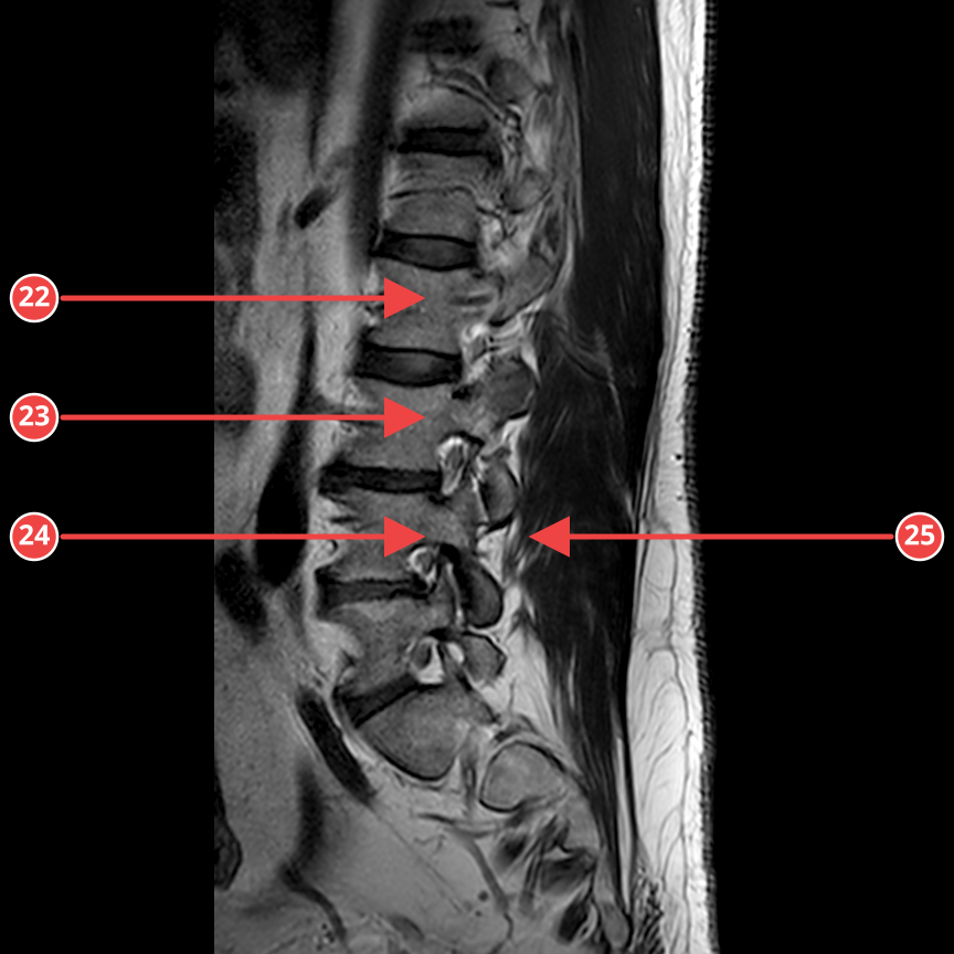
IMG-0002-00014.dcmT2_SAG
Sagittal T2-weighted sequence of the lumbosacral spine. A central L5-S1 herniation with compression of the dural sac and a small L4-L5 protrusion are visible. The remaining discs are within age-related norms.
Anomalies
- [1] L5-S1 disc herniation - high - Central herniation with compression of the dural sac, up to 5 mm. confidence 85%
- [2] L4-L5 disc protrusion - medium - Circular protrusion, most pronounced centrally, up to 3 mm. confidence 82%
- [3] L3-L4 disc degeneration - low - Decreased T2W signal, early signs. confidence 75%
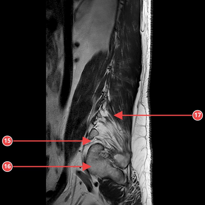
IMG-0002-00018.dcmT2_SAG
Sagittal T2-weighted sequence of the hip joint. A small amount of fluid in the joint cavity and heterogeneity of the articular cartilage are seen.
Anomalies
- [4] Hip joint effusion - medium - A moderate amount of free fluid in the joint cavity. confidence 75%
- [5] Articular cartilage heterogeneity - medium - Foci of thinning and signal heterogeneity of the cartilage in the weight-bearing zone. confidence 60%
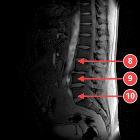
IMG-0001-00005.dcmSURVEY
Coronal survey view. Signs of degenerative changes of the intervertebral discs at several lumbar levels are visualised.
Anomalies
- [6] L4-L5 disc degeneration with protrusion - medium - Narrowing of the intervertebral space, decreased signal, protrusion. confidence 75%
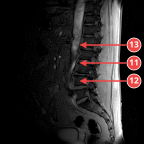
IMG-0001-00004.dcmSURVEY
Survey view with accentuated lordosis. Decreased signal of the L4-L5 and L5-S1 discs without significant compression.
Anomalies
- [7] Decreased L4-L5 disc signal - medium - Dehydration with moderately decreased T2 intensity. confidence 70%
Want the same report for your own images? Upload your study - processing takes 20-40 minutes.
Upload your studyPlease note: the AI result is for informational purposes only and does not replace a consultation with a doctor.