A detailed report
on your scans
Upload an MRI, CT, X-ray, or fluorography study - the AI will find anomalies, mark them with a red arrow directly on the images, and send a structured PDF report.
Just need to open an MRI disc? View your scan for free - no software to install.
Everything you need for a second opinion
From upload to a signed PDF - one flow with no extra steps.
Upload the entire study folder - the images are extracted automatically. Or pick individual JPG/PNG/DICOM files.
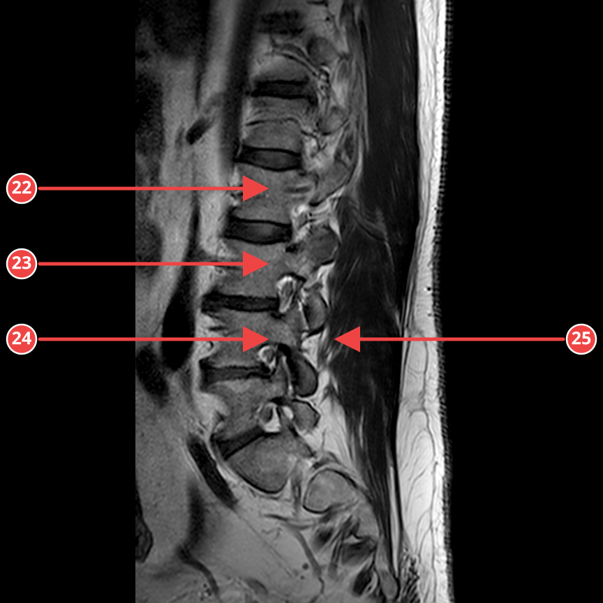
Every detected pathology is marked with an arrow directly on the image, with a number and coordinates, so it is easy to match with the report.
Structure: complaints and the examined area, a description of every image, an integrated conclusion, and recommendations for next steps.
Up to 500 images per run. For large studies the AI picks the key slices in each series itself, saving time and tokens.
Upload several studies from different dates in one batch - they are separated by DICOM tags, and the report gets a section on what changed and what stayed stable.
Files are processed in a secure environment and automatically deleted after the report is issued. Nothing is published.
Spine, head, chest, joints, abdomen. T1/T2/FLAIR/DWI support for MRI and standard projections for the other modalities.
How it works
Drag in individual images or a whole folder with DICOMDIR. The system picks the key images itself.
The service is in a free beta: enter the SABAHHUB promo code at the activation step - full functionality at no charge.
The neural network processes every image, highlights anomalies, and produces a single conclusion.
The finished document with annotated images and an interpretation can be downloaded or sent to your doctor.
The service is free for now
Beta mode: all plans are unlocked for free with the promo code SABAHHUB - enter it at the activation step.
- One study of up to 500 images
- Sampling: the AI analyzes representative slices
- Red arrows on anomalies
- PDF with a description and recommendations
- DICOMDIR and compressed format support
- Report access for 30 days
- Several studies at once - up to 1000 images in total
- Change analysis: what changed since last time and what is stable
- Analyzes every image - no sampling
- Premium Claude Opus model at maximum tokens
- Comparison table: "detected anomaly vs. norm"
- All images in the PDF with arrows on problem areas
- Model confidence for every finding
- Report access for 30 days
- API integration
- Branded PDF
- SLA and a private instance
- Dedicated manager
Frequently asked questions
What types of studies are supported?+
MRI, CT, X-ray, and fluorography. Spine, head, chest, joints, abdomen, and pelvis - any area where radiology imaging is used.
What file formats are supported?+
JPG, PNG, WebP, single DICOM files, and a full DICOMDIR folder - including compressed transfer syntaxes (JPEG, JPEG-LS, JPEG 2000, RLE). Key images are extracted from DICOMDIR automatically for each series.
How many images can I upload?+
The Basic plan - one study of up to 500 images. The In-depth plan - several studies at once, up to 1000 images in total. If you have more, write to mail@aict.az. With a large number of slices, the system groups them by series (T1/T2/FLAIR/DWI for MRI, axial/coronal for CT, projections for X-ray) and sends the key representative slices for analysis.
Can I compare two studies from different years?+
Yes, that is the In-depth plan. Upload both studies in one batch: they are separated automatically by DICOM tags, and the report gets a "Changes over time" section - what appeared, what grew or shrank, and what stayed stable. If the studies are not comparable (different areas or modalities), the report will say so directly instead of forcing a comparison.
Can I just open a scan without a report?+
Yes. On the "Open DICOM" page, a file from an MRI or CT disc opens in the browser for free, with no software to install: you can download the image as PNG and send it to your doctor, and the AI will describe what it shows. Free - up to 5 images.
What does the report look like?+
A PDF document: an introduction, a description of every image, an overall conclusion, and recommendations. Every image with an anomaly comes with a copy of the image marked with a red arrow.
Does this replace a doctor?+
No. The report is a second opinion and an aid for discussion with your attending physician, not a medical diagnosis.
Is my data safe?+
Files are encrypted and deleted after the report is generated - identifiable source images are not used for training. Only de-identified data that cannot be used to identify a patient may be used to improve model quality.
Is the service really free?+
Yes, everything is free while the beta lasts. Enter the SABAHHUB promo code at the activation step, and the analysis will start at no charge.
What language is the report in?+
The report is generated in three languages at once: first in Azerbaijani, then in Russian and English - all in one document.